Lipolytic enzymes are a group of enzymes that break down water-insoluble esters and produce fatty acids. They are used in many industrial processes including detergents, fuels, clothing, beverage and pulp. 

Phospholipase Enzymes

Phospholipase A2 (PLA2):

–Structure/mechanisms of action: PLA2 belongs to the lipolytic family of enzymes that hydrolyze the ester bond at the sn-2 position of the phospholipids. Upon hydrolysis of the phospholipids, PLA2s release free fatty acids and generate lysophospholipids (LPLs). Free fatty acids such as arachidonic acid (AA) and oleic acid (OA) are important sources of energy. (Khan, “The Phospholipase A2 Superfamily: Structure, Isozymes, Catalysis, Physiologic and Pathologic Roles” Intl J. Mol Sci, 24(2), 2023). 

A Ca2+ ion is required for both binding of the substrate and for catalysis. However, Group VI phospholipase A2 enzymes are members of the PLA2 superfamily that are characterized as Ca2+-independent PLA2 (iPLA2) enzymes. (Khan, “The Phospholipase A2 Superfamily: Structure, Isozymes, Catalysis, Physiologic and Pathologic Roles” Intl J. Mol Sci, 24(2), 2023).

PLA2s exist as active enzymes and as inactive PLA2-like proteins in snake venom. PLA2 enzymes catalyse the hydrolysis of the sn-2 ester bond of cell-membrane phospholipids and are classified into 14 groups, from which the groups IA and IIA are present in elapid and viperid venoms. (Fernandes, “The chemistry of snake venom and its medicinal potential” Nature. 6, 451-469 2022). pages451–469 (20pages451–469 (2022)

–Functions:

The PLA2s play important roles both physiologically and pathologically, with their expression increasing significantly in diseases such as sepsis, inflammation, different cancers, glaucoma, obesity and Alzheimer’s disease. Khan, “The Phospholipase A2 Superfamily: Structure, Isozymes, Catalysis, Physiologic and Pathologic Roles” Intl J. Mol Sci, 24(2), 2023).

The PLA2 superfamily has been classified into 16 groups (groups I to XVI), based on the chronology of their discovery, as well as based on their location in the body, substrate specificity. Khan, “The Phospholipase A2 Superfamily: Structure, Isozymes, Catalysis, Physiologic and Pathologic Roles” Intl J. Mol Sci, 24(2), 2023).

PLA2-IIA also plays a major role in host defense via its antimicrobial activity. It degrades bacterial membrane by hydrolyzing phosphatidylethanolamine and phosphatidylglycerol, which are abundant in the bacterial membrane. Blood plasma concentration of sPLA2-IIA can increase up to 500-fold in patients with acute diseases such as sepsis, peritonitis and bacterial infection, compared to healthy persons. (Khan, “The Phospholipase A2 Superfamily: Structure, Isozymes, Catalysis, Physiologic and Pathologic Roles” Intl J. Mol Sci, 24(2), 2023).

Secreted PLA2s, especially groups IIA, V and X, can play an important role in atherosclerosis. A study showed that young adults (age 24 to 39 years) with cardiovascular disease had an increased level of GIIA sPLA2 in their blood plasma, which may contribute to atherogenicity. (Khan, “The Phospholipase A2 Superfamily: Structure, Isozymes, Catalysis, Physiologic and Pathologic Roles” Intl J. Mol Sci, 24(2), 2023).

sPLA2s are widely known to be overexpressed in various tumors and cancer cells. (Khan, “The Phospholipase A2 Superfamily: Structure, Isozymes, Catalysis, Physiologic and Pathologic Roles” Intl J. Mol Sci, 24(2), 2023).

Metabolomics is the study of the entire complement of all the small molecular weight metabolitesinside an organism of interest. Some metabolites which are routinely assayed in the blood include urea for liver and kidney function, cholesterol for risk of coronary artery disease, and glucose. This can be a difficult task, however, because metobolites differ widely in their chemical characteristics (solubility, charge, MW, etc), and concentrations can differ by many orders of magnitude. 

The interaction of an organism with its environment is essential to its survival. The basis of this interaction is predominantly small molecules. On a molecular level, small molecules both promote, as in nutrients, and challenge, as in toxins, cell viability. Those gene products that interact with small molecules underlie the organism’s ability to adapt to environmental changes and include those that bind, transport, and metabolize small molecules. 

In the future, databases of metabolomic information will be used to establish the relations among metabolite profiles and health status. One will be able to compare the metabolic status of an individual by comparing the results of diagnostics to the reference knowledge base. This knowledge will allow interventions such as drugs to improve health and prevent disease. 

One can already see such an approach being used. For example, metabolic profiling of amino acids and acylcarnitines from blood spots by automated  is a diagnostic tool for errors of metabolism in newborns. 

Lipidomics is a branch of metabolomics in which non-water soluble metabolites are studied in relation to the function of genes and their proteins. Classical methods of lipid analysis utilize the techniques of thin layer chromatography (TLC), HPLC, gas liquid chromatography (GLC), and MS. The recently developed technique of  has allowed almost complete analysis of the lipidome from organic extracts of cells/tissues. 

The complete analysis of lipid metabolites has been applied to investigate the effects of TZDs which are therapeutic agents against type II diabetes. TZDs decrease serum lipid concentrations but these actions are accompanied by lipid accumulation in tissues. In a study by Watkins, an assessment of the lipid metabolome (the concentration of each lipid class and each of its constituent fatty acids) was applied to evaluate the effect of feeding a low does of a TZD on the lipid metabolisms of diabetic mice. Analysis of the results revealed key targets of the actions of the TZD.

Information used to predict genes includes signals in the sequence, content statistics and similarity to known genes. As noted by Attwood (Science, 290, 2000), there are many obstacles to accurate gene counting. First there is the problem of what exactly constitutes a gene. Is it a heritable unit corresponding to an observable phenotype? Is it a packet of genetic information that encodes an RNA or a protein? Must it be translated? Answeres to these question affect estimates of the total number of genes in sequenced genomes. 

Cross-References: Protein Bioinformatics DNA/RNA Bioinformatics

———————————————————————————————-

DNA Databases BLAST Genbank at NCBI EMBL DNA DDBJ

http://www.ncbi.nlm.nih.gov/LocusLink/” style=”color: rgb(146, 172, 186); -webkit-transition: background-color 0.2s ease-in-out, color 0.2s ease-in-out; transition: background-color 0.2s ease-in-out, color 0.2s ease-in-out;”>LocusLink (NCBI)

Annotation on genes: MedMiner

Analysis of complete genomes: PEDANT

Cloning/Enzymes: BRENDA SwissProt Restriction Enzyme Database Cloneit DNA artist download

Cut your DNA Sequence: NEB Cutter Web Cutter

Vector contamination: EMBL EBI NCBI VecScreen

Mammalian Gene Collection:  Mammalian Gene Collection (MGI) (cDNA sequences for human, rat & mouse) 

enes involved in Human Disease: GeneCards OMIN

Open Reading Frames (OPFs): ORF Finder ORF Finder (NCBI) Expasy-translate Sequence Manip Site

Comparison of genomes: Institute for Genomic Research TIGR Vista PipMaker

Sequence Submission: GenBank Submissions (BankIt) NCBI GenBank Examples NCBI Sequin (This is software developed by NCBI which must be downloaded. Use this for sequences which are too short to submit online with BankIt or where you want more control over your submissions) Sequin Help Sequin Factshttp://www.ebi.ac.uk/embl/Submission/webin.html” style=”color: rgb(146, 172, 186); -webkit-transition: background-color 0.2s ease-in-out, color 0.2s ease-in-out; transition: background-color 0.2s ease-in-out, color 0.2s ease-in-out;”>EMBL (WebIn)

Sequence Analysis: Metagene

Sequence Conversion: (Convert sequence files from one particular format to another format)

ReadSeq Readseqimple ReadseqBaylor Readseqfinice Readseqbioportal readseqebi Redseqbimas

Specific genomes: FlyBase newsnetwork

Wiring Diagrams of life: KEGG

Definitions:

Oxidation refers to where electrons are transferred from one atom to another. The molecule that gives up electrons is said to be Oxidized and is also called a reductant because it is reducing another molecule.

Reduction refers to where electrons are added. A molecule that accepts electrons is said to be an oxidant.

Nucleophile typically shares an electron pair with an electrophile in the process of bond formulation. In other words, a nucleophile is seeking a center of electron deficiency with which to react. Nucleophiles (“nucleus loving”) can be negatively charged or uncharged, and include for example heteroatoms other than carbon bearing a lone pair, or pi electrons in any alkene or alkyne.

Electrophiles (“electron-loving”) are electrically neutral or positively charged and have some place for the electrons to go, but it an empty orbital (as in BH3) or a potentially empty orbital.

Example: A common characteristic of a nucleophilic reaction that takes place on saturated carbon, is that the carbon atom is almost always bonded to a heteroatom, defined as an atom other than carbon or hydrogen. The heteroatom is usually more electronegative than carbon and is called the “leaving group” (L) in the substitution reaction. The leaving group departs with the electron pair by which it was originally bound to the carbon atom. The reactivity of a reactive group is largely determined by the tendency of a leaving group to depart. Another factor on reactivity of a reactive group is the strenght of the bond between the leaving group and the carbon atom, since this bond must break if substitution is to occur.The ease with which a leaving group departs is related to the basicity of that group.

Weak bases are in general good leaving groups because they are able to accommodate the electron pair effectively. Good leaving groups are in general the conjugate bases of strong acids with pKa values below 5 such as I-, Br-, Bl-. In general, a good leaving group is electronegative to polarize the carbon atom, it is stable with an extra pair of electrons once it has left, and is polarizable, to stabilize the transition state. With the exception of iodine, all the haolgens are more electronegative than carbon. A carbon-haologen (C-X) bond in an alkyl halide, for example, is polarized, with a partial positive charge on the carbon and a partial negative charge on the halogen. Thus the carbon atom is susceptible to attack by a nucleophile (a reagent that brings a pair of electrons) and the halogen leaves as the halide ion (X-), taking on the two electrons form the C-X bond.

Acids: as defined by the Bronsted/Lowry theroy of acid and bases are substances that donate hydrogen ions (H+).

Bases:  are substances that accept hydrogen ions.

Conjugate pairs: Examples of conjugate pairs include carboxylic acids which contain the carboxyl group -COOH. R-COOH is the conjugate acid and R-COO- is the conjugate base. A typical reaction would be R-COOH + H2O to yield R-COO- + H2O + H+.

pH lowering: The hydrogen ions which are released into the medium in the above reaction lowers the pH.

raising the pH: The removal of hydrogen ions or the addition of hydroxide ions (OH-) to water aises the pH.

Types of Bonds

(1) Hydrocarbons: C and H form covalent bonds. These hydrocarbons are nonpolar, do not form hydrogen bonds and are usually insoluble in water.

biobon1

(2) C- O bonds: play essential roles in carbohydrate metabolism and are also important in pharmaceutical products. For example, pharmaceutically desirable morphinan compounds which are extensively used for pain relief often have a ketone group (a keytone comprises “a carbonyl group” any oxygen atom double-bonded to a C atom where the C atom of the carbonyl group is single bonded to two carbon atoms.

  biobon2

(3) C-N bonds (These also occur in nucleic acids)

biobon4

(4) Phosphates

biobon5

(5) Disulfide bond: The formation of disulfide bonds from thiols of a protein is a two electron oxidation reaction requiring the presence of an appropriate electron acceptor (oxidant). Protein thiol oxidation to the disulfide is carried out by low molecular weight disulfide reagents, such as oxidized glutathione or cystamine. The cytoplasm of both eukaryotes and prokaryotes is highly reducing in nature, so the equilibrium constants for thiol disulfide exchange processes are unfavorable for the formation of disulfide bonds. In eukarytoes, the formation of disulfide bonds is more favorable in the secretory pathway because ER is significanlty oxidizing due to the lwo ration of GSH/GSSG, and PDI residues in the luen of the ER. In a gram negative bacteriu, the disulfide bond formation occurs in the periplasm with the help of DsbA. Thiol-disulfide exchange occurs via direct nucleophilic attack of the thiolate anion (S-_ on one of the sulfurs of the oxidant, forming a transient intermediate. As a result of nucleophilic substitution a sulfur atom is removed and a mixed disulfide intermediate is formed. The formation of a disulfide bond is favored (a) at pH of a 8-9 to favor the formation of the thiolate anions (S-_ and (b) when the two Cys-residues are close to each other.

(6) Covalent Bonds form when 2 atoms come very close together and share one or more of their electrons. In a single bond, one electron from each of the 2 atoms is shared (an example is H2 or H-H or hydrogen gas); in a double bond a total of four electrons are shared (example O2 or oxygen gass). Each atom forms a fixed number of covalent bonds in a defined spatial arrangement. C, for example, forms 4 single bonds arranged tetrahedrally.

Often electrons are shared unequally which results in a polar covalent bond. For example, the covalent bond between O and H, or N and H, is polar whereas that between C and H has electrons attracted much more equally by both atoms and is relatively nonpolar.

Noncovalent Attractions or Bonds

(1) Hydrogen Bonds: A hydrogen bond forms when a H atom is sandwiched between 2 electron attracting atoms (usually O or N). Hydrogen bonds are very important with respect to water. 2 atoms, connected by a covalent bond, may exert different attractions for the electrons of the bond. In such cases the bond is called “polar” with one end slightly negatively charged and the other end positively charged. This is exactly what happens with water. Although a water molecule has an overall neutral charge (the same # electrons and protons), the oxygen nucleus draws electrons away from the hydrogen nuclei, leaving the H nuclei with a small net positive charge. The small negative charge on the O atom can in turn hydrogen bond with a H atom on a neighboring water molecule.

biobon3Molecules that contain nonpolar bonds (CH bonds) are usually insoluble in water and are called “hydrophobic.” Substances which are polar can dissolve readily in water and are termed “hydrophilic.” Hydrogen bonding is also used between nucleotide bases and is common between amino acids in polypeptide chains.

Hydrogen bonding is also very important with respect to stabilizing proteins. Such hydrogen bonds can form (1) between atoms of 2 peptide bonds, (2) between atoms of a peptide bond and an amino acid side chain and (3) between 2 amino acid side chains.

(2) Van der Waals attractions: The electron clud around any nonpolar atom will fluctuate producing a flickering dipole. Such dipoles will transiently induce and oppositely polarized flickering dipole in a nearby atom. The 2 atoms will be attracted to each other in this way until the distance between their nuclei is about equal to the sum of their van der Waals radii. Although weak, these forces can become important when 2 macromolcecular sufaces fit very close together, because many atoms are involved.

(3) Ionic Bonds: occur when electrons are donated by one atom to another in contrast to covalent bonds which involve the sharing of electrons. Ionic bonds are more likely to be formed by atoms that have just 1 or 2 electrons in addition to a filled outer shell or are just one or 2 electrons short of acquiring a filled outer shell. They can often attain a completely filled outer electron shell more easily by transferring electrons to or from another atom than by sharing electrons. For example, Na, with atomic # 11, can strip itself down to a filled shell by giving up the single electron external to its second shell. By contrast, a Cl atom, with atomic number 17, can complete its outer shell by gaining just one electron. When an electron jumps from Na to cl, both atoms become electrically charged ions (Na+, Cl-). Because of their opposite charges they are attracted to each other and held together in an ionic bond.

When a sodium atom donates an electron to a chlorine atom, the soidum atom is oxidized and the chlorine atom is reduced. 

(4) Hydrophobic Force: This force is caused by a pushing of nonpolar surfaces out of the hydrogen bounded water network. The force is rather nonspecific but central to the proper folding of protein molecules.

See also Glycan Binding Proteins

Sialic acid (Sia)-binding Ig-like lectins (Siglecs) are a family of glycan-binding proteins (GBPs). They are an immunoglobulin super family lectins with a defining feature of recognizing sialic acid containing structures. Siglecs are I-type (Ig-type) lectins are are characterized by an N-terminal V-set Ig-like domain, which mediates Sia binding, followed by varying numbers of C2-set Ig-like domains.

Siglecs are cell surface receptors of the immunoglobulin super-family that regonize sialic acid containing proteins. Siglecs are primarily expressed by cells of hematopoietic origin. Most Siglecs are considered to be negative regulators of the immune system becasue they contain one or more cytosolic immune receptor tyrosie-based inhibitory motifs (ITIMs).

Families of Siglecs

There are two families of siglecs.

(I) Evolutionary conserved Siglecs: include the siglecs, Siglec-1, -2, -4, and -15. 

(II) Evolving CD33-related Siglecs: include CD33rSiglecs, Siglec-3, 5-14 and 16 in primates, and Siglec 3 plus E-H in rodents.

Functions

The primary biological functions of CD33rSiglecs remain unkown. One possibility is that they are self recognition molecules that prevent inappropriate activaiton of innate immune cells. An alternative hypothesis is that when unmasked they serve as detectors of invasive Si-expressing bacterai. Several strains of pathogenic bacteria are known to express Neu5Ac on their surfaces., thereby mimicking host cell surfaces and evading detection by both innate and adaptive immune systems. Unmasked Siglecs could thus provide a mechanisms for the innate immune system cells to recognize these camouflaged bacterai. Si-expressing pathogenci bacterai isolated form multiple mammalian species express Neu5Ac but never Neu5Gc. Thus, the pathogen recognition hypothesis predicts that CD33rSiglecs on cells of the innate immune system must recognize Neu5Ac. (Sonnenburg, Glycobiology, 14(4), 339-346, 2004).

Siglec-10 – CD24 interaction: It has been suggested that interaction of Siglec-10 with CD24 protects the host against a lethal response to pathological cell death by discriminating a danger-associated molecular pattern (DAMP) and repressing damage-induced immune responses during a noninfectious inflammatory reactions (Chen et al. 2009). It was discovered that interactions between Siglec-10 and CD24 mediated recognition of “self,” and thus limited damage of host cells by innate immune cells during a noninfectious inflammatory process, in which endogenous DAMPSs such as HMGB1 were involved in a complex with CD24 and Siglec-10. It is likely that the interactions between CD24 and Siglec-10 is between the sialic acid binding site of Siglec-10 and the heavily sialylated glycans of CD24 (Varki, Glycobiology, 19(8) (2009)).

IgGglycosylationIgGglycoforms

Raju “Glycosylation Variations with Expression Systems and their impact on biological activity of therapeutic immunoglobulins” BioProcess International, April 2003

Fig. 4 above shows the largest N-linked oligosaccharide structure found in human IgG. This structure is present as a very minor component. The majority of oligosaccharides found in human IgG are truncated versions of the structure shown in Fig. 4. The microheterogeneity of human IgG glycans is mainly due to the presence and/or the absence of eitehr core fucose, bisecting GlcNAc, termial sialic acid, exposed Gal, and often the exposed GlcNAc residues. See Raju above 

In human IgG1, which is the main subtype used for therapeutics, the majority of the Fc glycans are complex biantennary structures wtih variable galactosylation: 1, 1, and 2 terminal galactoses corresponding to G0, G1 and G2 glycoforms, respectively. (Boeggeman, Bioconjugate Chem. 2009, 20, 1228-1236).

G0 glycan: refers to the complex N-linked glycan having the G1cNAc2Man3G1cNAc2 core structure, where no terminal sialic acids (NeuAcs) or terminal galactose (Gal) sugar residues are present. In IgG-GO glycoforms, neither arm is galactosylated so that a GlcNAc residue is exposed at the terminals of each arm. About 20% of the IgG glycans at this glycosylation site terminate in GlcNAc on both arms The IgG-G0 glycoforms have been shown to bind MBL. (Arnold, J. Immunol, 2004 173: 6831-6840). 

G1 glycan: refers to the complex GNGN biantennary N-glycan having the G1cNAc2Man3G1cNAc2 core structure plus one terminal galactose residue. In IgG-G1 glycoforms, a terminal galactose residue is missing from one arm, exposing a GlcNAc residue. (Arnold, J. Immunol, 2004 173: 6831-6840)

G2 glycan: refers to complex GNGN biantennary N-glycan having the G1cNAc2Man3G1cNAc2 core structure plus two terminal galactose residues. (Arnold, J. Immunol, 2004 173: 6831-6840) 

Production of Immunoglobuilin Glycoforms

G0 glycoforms: Raju (US2007/0041979) discloses preparation of G0 glycoform by treating IgG samples with a sialidase A to remove minor amounts of terminal sialic acid residues followed by treating with beta galactosidase to remove terminal beta-galactose residues. After purificaiton on a protein A column, the resulting G0 glycoform was characterized by MALDI-TOF-MS.

Fox (US2010/0266607) discloses an antibody glycoform in which sialic acid and terminal beta galactose residues are removed by incubation with sialidase and beta galactosidase.

G1 glycoforms: (Hueller, US14348822) discloses a method for producing an immunoglobuilin with a G1 glycostructure from a mixture of a glycosylated antibody or fusion polypeptides of the G0, G1 and G2 glycostructures. The method involves the following sequences of events 1) incubate with a galactosyltransferase, 2) incubate this product with a sialytransferase, 3) incubate this product with a beta-1,4-galactosidase to oobtain a beta-1,4-galactosidase reaction product, 4) remove or inactive the beta-1,4-galactosidase and 5) incubate the reaction product with a sialidase. 

G2 glycoforms: Raju (US2007/0041979) discloses preparation of G2 glycoform by treating IgG samples with a sialidase A, purifying on Protein A and then treating the sialidase A IgG samples with beta 1, 4 GT (including UDP-Gal) for 24 hours.

Fox (US2010/0266607) discloses an antibody glycoform which is maximally sialyated by incubation of antibody with beta 1,4-galactosyltransferase and alpha 2,3-sialytransferase and UDP-galactose, CMP-N-acetylnueraminic acid and MnCl.

Free of Core Fucoses: (Wong, US2011/0263828) discloses methods for modifying the Fc region glycosylation pattern of an antiboyd by providing a clycosylated Fc region, contacting the glycosylated Fc region with alpha-fucosidase to remove core fucose residues, contacting the de-fuosylated Fc with beta-1,4-galactosyltransferaseand then alpha-2,6-sialytransferase, then with beta-1,4 galactosyltransferase under conditions suitable for attaching a galatose to the oligosaccharide and then contacting the Fc region with alpha-2,6-sialytransferaseunder conditions suitable for attaching a terminal sialyl residue. 

Free of Galactose:

Raju (US2010/0260751) discloses that host cells may be devoicd of galactosyltransferases such that antiboides expressed in these cells lack galactose. Wihtout galactose, sialic acid will also not be attached.

–Removal of (alpha 1,3) glycosidically bound terminal galactose residues):  diclsoes that alpha 1,3 galactosidases from green coffee beans (EC 3.2.1.22) can be used to selectively remove (alpha 1,3) glycosidically bound terminal galactose residues from the oligosaccharide attached at amino acid residue Asn297 to an immunoglobuiolin CH2 domain.


See also determining ABO Blood type   See also galactosidases   See removing A/B antigens from blood cells

Blood group antigens are either sugars or proteins, and they are attached to various components in the red blood cell membrane. For example, the antigens of the ABO blood group are sugars. In contrast, the antigens of the Rh blood group are proteins.ygotes of IAi heterozgotes.

2. T

The ABO blood group system was the first to be discovered and is the one of greatest importance from the standpoint of blood transfusion. Individuals of blood types A, B and O express A, B and H antigens respectively. These antigens are found on the surface of erythrocytes and platelets as well as on the surface of endothelial and epithelial cells. The major blood product used for transfusion is erythrocytes, which are red blood cells containing hemoglobin. The blood group structures are glycoproteins or glycolipids.

The ABO blood group specificity is determined by the nature and linkage of monosaccharides at the ends of the carbohydrate chains. The carbohydrate chains are attached to a peptide (glycoprotein) or lipid (glycosphingolipid) backbone, which are attached to the cell membrane of the cells. The immunodominant monosaccharide determining type A specificity is a terminal alpha1-3 linked N-acetylgalactosamine (GalNAc), while the corresponding monosaccharide of B type specificity is an alpha1-3 linked galactose (Gal)Type O cells lack either of these monosaccharides at the termini of oligosaccharide chains, which instead are terminated with alpha1-2 linked fuctose (Fuc) residues.

Blood of group A contains antibodies to antigen B. Conversely, blood of group B contains antibodies to antigen A. Blood of group AB has neither antibody and blood group O has both. An individual whose blood contains either (or both) of the anti-A or anti-B antibodies cannot receive a transfusion of blood containing the corresponding incompatible antigen(s). If an individual receives a transfusion of blood of an incompatible group, the blood transfusion recipient’s antibodies coat the red blood cells of the transfused incompatible group and cause the transfused red blood cells to agglutinate. In order to avoid red blood cell agglutination, transfusion reactions, and hemolysis, transfusion blood type is cross matched against the blood type of the transfusion recipient. For example, a blood type A recipient can be safely transfused with type A blood, which contains compatible antigens.

Because type O blood contains no A or B antigens on its RBC membranes, it can be transfused into any recipient with any blood type. People with bloods types A, B, and AB have no antibodies to reject type O. Type O blood is often referred to as the “universal donor” for this reason.

In an attempt to increase the supply of type O blood, methods have been developed for converting certain type A, B and AB glood to type O blood using galactosidases which enzymatically remove the immunodominant monosaccharides specifying the blood group A and B antigens. ygotes.

Gene Organization of teh ABO blood groups:

Mendel always looked at genes with two alternative alleles. Although any dipoloid inividual can carry only two alleles for a gene, there may be more thna two alleles in a population. The ABO blood types in humans invovles three alleles. 

The gene that determines ABO blood types encodes an enzyme that adds sugar molecuels to proteins on teh surface of red blood cells. These sugars are in trn recognition markers for the immune system.

The gene that encodes the enzyme, designated I, has three common alleles: IA, whose product adds galactosamine, IB, whose produce adds galactose, and i, which codes for a protein that does not add a sugar. The gree alleles of the I gene can be cominbed to produce six different genotpyes. An individual heterozygous for the IA and IB alleles produces both forms of the enzyme and exhibits both galactose and galactosamine on red blood cells. Because both alleles are expressed simultaneously in heterozygotes, the IA and IB alleles are condominant. Both IA and IB are dominant over the i allele, becasue both IA and IB alleles lead to sugar addition, whereas the i allele does not. The different combinations of the three alleles produce the following four different phenotypes:

(1) Type A inidivduals add only galactosamine. They are either IAIA homozgotes or IAi heterozyals add only galactose. 

(2) Type B individuls add only galactose. They are either IBIB homozygoes or IBi heterozygotes.

(3) Type AB individuals add both sugars and are IAIB heterozgotes.

(4) Type O individuals add neither sugar and are ii homozygotes. 

The Specific Antigens on A, B and O RBCs

Individuals of blood types A, B and O express A, B and H antigens respectively. These antigens are not only found on the red cells but on the surfaces of all endothelial and most epithelial cells as well.

N-Acetylgalactosamine (GalNAc): is an amino sugar derivative of galactose. It is the terminal carbohydrate forming the antigen of blood group A. It is typically the first monosaccharide that connects serine or threonine in particular forms of protein O-glycosylation.

ABH Antigen Subgroups

The ABH antigens are divided into subgroups, depending on the inner core saccharide chain. As an example, both A, B and H antigens are expressed on type 1 (Galbeta1, 3 GlcNAc), type 2 (Galbeta1,4GlcNAc), type 3 (Galbeta1,3GalNAcalpha) and type 4 (Galbeta1,3GalNAcbeta) chains. (Holgersson (US8,404,456).

Lectins are glycoproteins that can bind carbohydrates. These carbohydrate binding proteins serve a number of vital roles which are disucussed below.

Classification

There are a number of structurally distinct plant and animal lectin families. Lectins have been separated into various classes based on what type of carbohydrate they bind to. Schnorr (WO2004/090549). 

C-type lectins (selectins): There are 32 extracellular C-type lectins (selectins)

–Mannose-binding lectin (MBL) and ficolin: These proteins belong to the C-type lectin family (or collecitns) are are ocmposed of multiple subunits containing cysteine and collagen rich domains, an alpha helical coil and a highly conversed carboxy-temrinal carohydrate recognition domain (CRD). These lectins activate the lectin pathway of the complement system. 

I type (or sialic aicd) lectins: There are two I type (or sialic aicd) lectins that have been characterized. 

Biological Functions

(1) intracellular sorting: An example of this is lysosomal hydolases which are transported through the golgi and eventually end up in lysosomes. These hydrolases have a unique marker, Mannose-6-phosphate, which are recognized by M6P receptors in the trans-golgi.

(2) clearance of serum proteins; sialic acids on the nonreducing end of oligosccharide chains protects those proteins from rapid degradation in the serum. Exposure of galactose after removal of sialic acid render the protein susceptible to the binding of a receptor on hepatocytes which take up the complex by endocytosis and degrade it in lysosomes. Most peptide and proteins that are injected into humans are also rapidly degraded unless carbohydrate is attached. Proteins with manose or galactose as the nonreducing terminal sugar are rapidly recognized by mannose or galactose receptors. Pharmaceutical companies likeNeose are developing strategies to glycosylate proteins to improve serum half life, using chemical and enzymatic techniques.

(3) complement cascade; Mannose binding lectins found in the serum bind 3 adjacent mannose residues on the surface of invading pathogens which helps initiate the complement cascade. Host structures do not have 3 mannose residues this close together, and are not recognized. Mannose-binidng lectin (MBL) and ficolin, an MBL-like protein, activate the lecitn pathway of the complement system. These proteins belong to the C-type lectin family (or collectins) and are composed of multimple subunits containing cysteine and collagen-rich domains, an alpha helical coil and a highly conserved carboxy-terminal carbohydrate recognition domain (CRD). See the complement system for more detail. 

(4) cell-cell interaction and inflammation; The endothelium express which upon binding to their carboyhydrate containing receptors on neutrophils, initiate a rolling adhesion of neutrophils. The neutrophils in turn activate their integrins which bind to endotheilial ICAM permitting a firmer adhesion.

(5) dendritic cell function: It has been demonstrated that lectins function in dendritic cells to capture and direct antigens to specialized antigen-processing compartments within the cell, via receptor mediated endocytosis. Also, lectin-ligand interactions can modulate cytokine production by dendritic cells as well as the maturation state of those cells. One class of lectins that often appear are caclium dependent carbohydrate binding proteins, or “C-type lectins”. 

Applications

Companies like Glycominds have developed glycochips which allow detection of proteins or cells to particular oligosaccharide structures. These chips may prove useful for development of drugs that inhibit cell-cell interaction, such as occurs during inflammation.

As to purification of glycosylated antibodies see “antibody purification” and “glycan antibody” under “particular types”

See also Function of Glycosylation:  (see outline)

glycosylation 1  from WO 2007/024743 A2

antibodyglycosylation 1from US 14/352,411

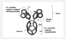
Antibodies are glycoproteins containing between 3 and 12% carbohydrate. The carbohydrate units are transferred to acceptor sites on the antibody chains after the heavy and light chains have combined. The major carbohydrate units are attached to amino acid residues of the constant region of the antibody. Carbohydrate is also known to attach to the antigen binding sites of some antibodies and may affect the antibody-binding characteristics by limiting access of the antigen to the antibody binding site. There are a number of roles associated with the carbohydrate units. They may affect overall solubility and the rate of catabolism of the antibody. It is also known that carbohydrate is necessary for cellular secretion of some antibody chains. It has been demonstrated that glycosylation of the constant region plays a vital role in the effector functioning of an antibody; without this glycosylation in its correct configuration, the antibody may be able to bind to the antigen but may not be able to bind for example to macrophages, helper and suppressor cells or complement, to carry out its role of blocking or lysing the cell to which it is bound (EP481790).

An integral feature of all normal IgG class antibodies is the N-linked oligosaccharides in the CH2 domain. The glycans most commonly associated with human serum IgG are complex bi-antennary chains N-linked to Asn-297 of the ?-H-chain. These branched sugar chains are situated within a cleft formed by the paired heavy chains in the CH2 domain such that they may undergo extensive non-covalent interactions with the adjacent polypeptide. Crystallographic studies on immunoglobulin Fc fragments have shown that the two CH2 domains do not form extensive lateral associations. The resultant interstitial region is filled by the inclusion of the oligosaccharide side chains attached to Asn-297 on each heavy chain such that the carbohydrates form a bridge across the domains. The alpha (1-6) arms of the biantennary complex oligosaccharides interact with the protein and the alpha (1-3) arms of the oligosaccharides form the bridge (Leatherbarrow, Molecular Immunology, 22(4), 1985).

Monoclonal antibodies are complicated glycoproteins subject to glycosylation heterogenicity and other modifications such as N-terminal pyroglutamate and C-terminal lysine variants, methione oxidation, asparagine deamidation, disulfide bond scrambling, aggregation, and fragmentation. Human serum IgG carries, on average, 2.8 N-linked oligosaccarides, of which 2.0 are invariably located in the Fc at the conserved N-glycosylation site of Asn 297. The additional oligosaccharides are located in the variable region of the light and heavy chains, with a frequency and position dependent on the occurrence of the N-glycosylation sequon (Asn/Xaa/Ser(Thr).

About 30 different biatennary oligoscaccharides are found to be associated wtih total human serum IgG. These are distributed non-randomly between the Fab and Fc. This heterogeneity creates a very large number of variants (glycoforms) of each unique IgG polypeptide causing further structural, and perhaps functional, diversification ((Rademacher, Springer Semin Immunopathol, 10, 1988, 231-249).

The oligoscaccharides present in an IgG antibody are complex and highly heterogeneous due to the presence or absence of various sugar risidues, including sialic acid, galactose (Gal), N-acetylglucosamine (GlcNAc), bisecting GlcNAc, and the core fucose (Fuc residues). (Qian, Anal. Biochem. 364 (2007) 8-18

Glysocylation at Position 297 (Asn 297):

Human immunoglobulins are mainly glycosylated at the asparagine residue at position 297 (Asn 297) of the heavy chain CH2 domain with a core fucosylated biantennary complex oligosaccharide. The biantennary glycostructure can be terminated by up to two consecutive galactose (Gal) residues in each arm. The arms are denoted (1,6) and (1,3) accoridng to the glyscoside bond to the central mannose residue. The glycostructure denoted as G0 comprises no galactose residue, G1 contains one or more galactose residues in one arm, G2 contains one or more galactose residues in each arm. IgG contains a single, N-linked glycan at Asn297 in the CH2 domain on each of its two heavy chains. The coavlently linked, complex carbohydrate is compoased of a core, biantennary penta-polysaccharide containing N-acetylglucosamine (GIcNAc) and mannose (man). Further modification of the core carbohydrate structure is observed in serum antibodies with the presence of fucose, branching GIcNAc, galactose (gal) and terminal sialic acid (sa) moieties variably found. Over 40 different glycoforms have been detected to be covalently attached to this single glycosylation site (WO2008/057634). 

The carbohydrate is sequestered between the heavy chains and has a complex biantennary structure composed of a core saccharide structure consisting of two mannosyl residues attached to a mannosyl-di-N acetylchitobiose unit. The outer arms arise from the terminal processing of the oligosaccharide in the Golgi; although the overall structure of the carbohydrate is conserved, considerable heterogeneity is seen in the identity of the terminal sugar residues. Analysis of carbohydrates isolated from normal human serum IgG has yielded up to 30 different structures (WO 2006/088447, p. 46, 3rd ¶). IgG antibodies depleted of asparagine-linked carbohydrate chains loose the ability to activate complement, to bind Fc receptors on macrophages and to induce antibody-dependent cellular cytotoxiciy (Nose, Proc. Natl. Acad. Sci. USA, 80, 1983, 6632-6636). 

Galactosylation:

In human IgG1, which is the main subtype used for therapeutics, the majority of the Fc glycans are complex biantennary structures wtih variable galactosylation: 1, 1, and 2 terminal galactoses corresponding to G0, G1 and G2 glycoforms, respectively. (Boeggeman, Bioconjugate Chem. 2009, 20, 1228-1236).

Fucosylation:

Fucosylation refers to the pressence of fucose residues within the oligosaccharides attached to the peptide backbone of an antibody. Specifically, a fucosylated antibody includes alpha(1,6)-linked fucose at the innermost N-acetylglucosamine (GlcNAc) residue in one or both of the N-linked oligosaccharides attached to the antibody Fc region (e.g., at position Asn 297 of the human IgG1 Fc domain). (Freimoser-Grundshober, US 14/352411).


Serine protease inhibitors or “serpins” form a large family of inhibitors that regulate various physiological processes such as angiogenesis, neoplasmia, apoptosis, inflammation, coagulation, fibrinolysis and viral pathogenesis. Dysfunction of such serine protease inhibitors has been implicated in cancer, thrombosis, emphysema, cystic fibrosis, liver cirrhosis, psoriasis, arthritis, immune hypersenstivity, and mental disorders.

Some of the best characterized serpins, including alpha1-proteinase inhibitor, alaph1-antichymotrypsin, antithrombin III and alpha2-antiplasmin are inhibitors of serine proteinases. The inhibitory serpins regulate proteolysis by forming tight, equimolar complexes with a target proteinase. Once formed, the complexes are rapidly removed form the circulcaton by cellular receptors, thereby preventing proteoysis of blood and tissue proteins. Two distinct pathways for the plasma elimination of serpin complexes are a receptor designated serpin receptor 1 (SR1) which recognizes complexes of proteinases with alpha1PI, alpha1ACT, ATIII and herparin cofactor II. Similarly, SR2 recognizes and eliminates complexes of proteinases with alpha2AP. These receptors are mainly present on hepatocytes and are probably responsible for the bult removal of proteinase activity during episodes of inflammation, coagulation, and fibrinolysis. (Mast, Biochemistry, 1991, 30, 1723-1730).

Alpha 1-Antitrypsin (A1AT)(AAT)/ alpha-1 proteinase inhibitor (A1PI): is a serpine protease inhibitor belonging to the serpin superfamily. It is a glycoprotein which consists of a single polypeptide chain to which several oligosaccharide units are covalently bonded. Human alpha-1 proteinase inhibitor has a role in controlling tissue destruction by endogenous serine proteinases. It protects tissues form enzymes of inflammatory cells such as neutrophil elastase. In its absence, neutrophil elastase is free to break down elastin, which contributes to the elasticity of lungs, and can result in respiratory problems like emphysema or COPD. ?1-antitrypsin is the predominant elastase inhibitor in tissues whereas SLPI is the predmoninant elastase inhibitor in secretions. Inherited deficiency of A1AT predisposes individuals to early onset hereditary emphysema, due to the upregulated action of neutrophil elastase. Large quantities and frequent injection sof A1At are required to restore normalcy to the lungs and relieve hereditary emphysema. Presently, A1AT supplies are isolated from pooled blood products (e.g., Zemaira®, Prolastin®)., and there is a greater demand for the product relative to the available supply. A1AT may also be useful in the treatment of emphysema cuased by smoking, cystic fibrosis, pulmonary hypertension, pumonary fibrosis and CPOD.

Alpha 1-antichymotrypsin: is a serine protease inhibitor synethesized in the liver with apparent affinity toward chymotrypsin like enzymes. It is structurally related to AAT and may be useful in the treatment of pulmonary inflammation (US 4,916,117)

Alpha-2-Macroglobulin: is a large plasma protein found in blood and produced by the liver. It is composed of four identical subunits bound together by disulfide bonds. It is able to inactivate a wide variety of proteinases including serine-, cysteine-, aspartic- and metalloproteinases. It also functions as an inhibitor of coagulation by inhibiting thrombin. Alpha2M is synthesiezed as a precursor having 1474 amino acid residues. The first 23 amino acids function as a signal sequence that is cleaved to yield a mature protein with 1451 amino acid reisudes. Alpha2M promiscuously binds to proteins and peptides with nucleophilic amino acid side chains in a covalent manner. Protease interaction with alpha2M results in a complex structural rearrangement called transformation, which is the result of a cleavage within the “bait” region of alpha2M after the proteinase becomes trapped by theioesters. The conformational change exposes residues required for receptor binding, allowing the alpha2M-proteinase complex to bind to the alpha2MR.

Inter-alpha-trypsin inhibitor (ITI): is also known as inter-alpha (globulin) inhibitor. ITI is a high molecular mass glycoprotein (about 240 kDa) present in human plasma. It was first isolated in 1962. However, its function as a proteinase inhibitor was not established until 1965. When ITI is exposed to proteolytic enzymes (e.g., pancreatic trypsin or elastase), a low molecular mass compound with antiroteolytic activity, termed HI-30 appears in the reaction mixture. The molecular mass of HI-30 varies in the range between 30 and 60 kDa. The amino acid sequencing of HI-30 is identical to the acid-soluble serum-trypsin-inhibitor (STI) which appears in urine after its renal filtration and is therefore also named urinary-trypsin-inhbitor (UTI). UTI and STI have identical amino acid sequences but they may be different in their carbohyrate composition.

The I?I family in mammals refers to a group of related plasma protease inhibitors. These proteins are comprised of various multi-polypeptide molecules showing different assemblies from a group of four distinct polypeptides including 3 related heavy (H) chains H1, H2 and H3, and a light chain called bikunin. Two Kunitz-type protease inhibitor domains are present in the bikunin chain which are bound to the heavy chains through glycosaminoglycan bridges. In more detail, ITI consists of a light chain (L) serine protease inhibitor, known as bikunin or urinary trypsin inhibitor (UTI) and one or two heavy chains (H) containing von-Willebrand type A (vWA) domains. There are 5 known IAI heavy chain polypeptides, which are encoded by inter-alpha (globulin) inhibitor (ITI)H1, ITIH2, (ITIH3), ITIH4, ITIH5: The ITIs comprise a family of protease inhibitors found in the extracellular matrices of various organs, as well as in the blood circulation. Due to their isolation in complexes with hyaluranan (HA), ITIs are also referred to as SHAPs (Serum-derived HA-associated proteins). It has been shown that interaction of ITIs with HA leads to stabilization of the extracellular matrix. (Veeck, Ocogene, 27, 2008, 865-876). While IaI is most abundantly found in the liver, it is also present in the lungs.

 Plasminogen Activator Inhibitor Type-1 (PAI-1): is a serine protease inhibitor. PAI-1 is a fast-acting inhibitor of tissue-type plasminogen activator (t-PA) and urokinase-type plasminogen activator (u-PA). Studies have shown that locally elevated PAI-1 plasma levels are important for sucessor failure of thrombolytic therapy with rt-PA. For example, one study showed that TNK-t-PA which is 90 times more resistant to PAI-1 compared with wild type t-PA was also 7.5 times more potent than t-PA in lysing whole blood clots. The clinical relevance of these studies suggest that a PAI-1-resistant variant of t-PA should be used as a more potent fibrinolytic agent for thromboytic therapy as in myocardial infarction.

Send an Email. All fields with an * are required.