See also Function of Glycosylation: (see outline)
 from WO 2007/024743 A2
 from US 14/352,411
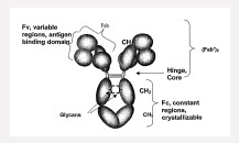
Antibodies are glycoproteins containing between 3 and 12% carbohydrate. The carbohydrate units are transferred to acceptor sites on the antibody chains after the heavy and light chains have combined. The major carbohydrate units are attached to amino acid residues of the constant region of the antibody. Carbohydrate is also known to attach to the antigen binding sites of some antibodies and may affect the antibody-binding characteristics by limiting access of the antigen to the antibody binding site. There are a number of roles associated with the carbohydrate units. They may affect overall solubility and the rate of catabolism of the antibody. It is also known that carbohydrate is necessary for cellular secretion of some antibody chains. It has been demonstrated that glycosylation of the constant region plays a vital role in the effector functioning of an antibody; without this glycosylation in its correct configuration, the antibody may be able to bind to the antigen but may not be able to bind for example to macrophages, helper and suppressor cells or complement, to carry out its role of blocking or lysing the cell to which it is bound (EP481790).
An integral feature of all normal IgG class antibodies is the N-linked oligosaccharides in the CH2 domain. The glycans most commonly associated with human serum IgG are complex bi-antennary chains N-linked to Asn-297 of the ?-H-chain. These branched sugar chains are situated within a cleft formed by the paired heavy chains in the CH2 domain such that they may undergo extensive non-covalent interactions with the adjacent polypeptide. Crystallographic studies on immunoglobulin Fc fragments have shown that the two CH2 domains do not form extensive lateral associations. The resultant interstitial region is filled by the inclusion of the oligosaccharide side chains attached to Asn-297 on each heavy chain such that the carbohydrates form a bridge across the domains. The alpha (1-6) arms of the biantennary complex oligosaccharides interact with the protein and the alpha (1-3) arms of the oligosaccharides form the bridge (Leatherbarrow, Molecular Immunology, 22(4), 1985).
Monoclonal antibodies are complicated glycoproteins subject to glycosylation heterogenicity and other modifications such as N-terminal pyroglutamate and C-terminal lysine variants, methione oxidation, asparagine deamidation, disulfide bond scrambling, aggregation, and fragmentation. Human serum IgG carries, on average, 2.8 N-linked oligosaccarides, of which 2.0 are invariably located in the Fc at the conserved N-glycosylation site of Asn 297. The additional oligosaccharides are located in the variable region of the light and heavy chains, with a frequency and position dependent on the occurrence of the N-glycosylation sequon (Asn/Xaa/Ser(Thr).
About 30 different biatennary oligoscaccharides are found to be associated wtih total human serum IgG. These are distributed non-randomly between the Fab and Fc. This heterogeneity creates a very large number of variants (glycoforms) of each unique IgG polypeptide causing further structural, and perhaps functional, diversification ((Rademacher, Springer Semin Immunopathol, 10, 1988, 231-249).
The oligoscaccharides present in an IgG antibody are complex and highly heterogeneous due to the presence or absence of various sugar risidues, including sialic acid, galactose (Gal), N-acetylglucosamine (GlcNAc), bisecting GlcNAc, and the core fucose (Fuc residues). (Qian, Anal. Biochem. 364 (2007) 8-18
Glysocylation at Position 297 (Asn 297):
Human immunoglobulins are mainly glycosylated at the asparagine residue at position 297 (Asn 297) of the heavy chain CH2 domain with a core fucosylated biantennary complex oligosaccharide. The biantennary glycostructure can be terminated by up to two consecutive galactose (Gal) residues in each arm. The arms are denoted (1,6) and (1,3) accoridng to the glyscoside bond to the central mannose residue. The glycostructure denoted as G0 comprises no galactose residue, G1 contains one or more galactose residues in one arm, G2 contains one or more galactose residues in each arm. IgG contains a single, N-linked glycan at Asn297 in the CH2 domain on each of its two heavy chains. The coavlently linked, complex carbohydrate is compoased of a core, biantennary penta-polysaccharide containing N-acetylglucosamine (GIcNAc) and mannose (man). Further modification of the core carbohydrate structure is observed in serum antibodies with the presence of fucose, branching GIcNAc, galactose (gal) and terminal sialic acid (sa) moieties variably found. Over 40 different glycoforms have been detected to be covalently attached to this single glycosylation site (WO2008/057634).
The carbohydrate is sequestered between the heavy chains and has a complex biantennary structure composed of a core saccharide structure consisting of two mannosyl residues attached to a mannosyl-di-N acetylchitobiose unit. The outer arms arise from the terminal processing of the oligosaccharide in the Golgi; although the overall structure of the carbohydrate is conserved, considerable heterogeneity is seen in the identity of the terminal sugar residues. Analysis of carbohydrates isolated from normal human serum IgG has yielded up to 30 different structures (WO 2006/088447, p. 46, 3rd ¶). IgG antibodies depleted of asparagine-linked carbohydrate chains loose the ability to activate complement, to bind Fc receptors on macrophages and to induce antibody-dependent cellular cytotoxiciy (Nose, Proc. Natl. Acad. Sci. USA, 80, 1983, 6632-6636).
Galactosylation:
In human IgG1, which is the main subtype used for therapeutics, the majority of the Fc glycans are complex biantennary structures wtih variable galactosylation: 1, 1, and 2 terminal galactoses corresponding to G0, G1 and G2 glycoforms, respectively. (Boeggeman, Bioconjugate Chem. 2009, 20, 1228-1236).
Fucosylation:
Fucosylation refers to the pressence of fucose residues within the oligosaccharides attached to the peptide backbone of an antibody. Specifically, a fucosylated antibody includes alpha(1,6)-linked fucose at the innermost N-acetylglucosamine (GlcNAc) residue in one or both of the N-linked oligosaccharides attached to the antibody Fc region (e.g., at position Asn 297 of the human IgG1 Fc domain). (Freimoser-Grundshober, US 14/352411).

